How to Stage Myxomatous Mitral Valve Disease
and Feline Cardiomyopathy with
2D Echocardiography
Canine MMVD
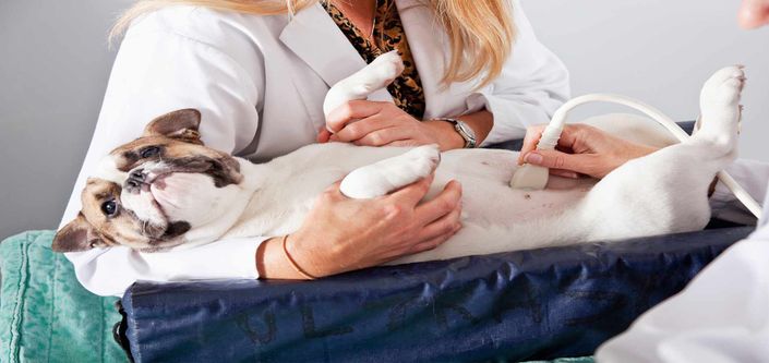
Echocardiography is the most sensitive method to measure heart size and is now the diagnostic test with highest priority in dogs with preclinical MMVD.
This course will teach you how to acquire and accurately measure the left atrium and left ventricle using simple right parasternal 2D echocardiographic images allowing you to diagnosis Stage B2 MMVD and recommend pimobendan treatment according to the 2019 ACVIM MMVD Consensus statement guidelines.
Feline Cardiomyopathy
The best method to discriminate pathologic from non-pathologic murmurs is echocardiographic assessment with emphasis on accurate assessment of left atrial size.
This course will teach you how to acquire and accurately measure the left atrium using simple right parasternal 2D echocardiographic images allowing you to stage the severity of cardiomyopathy.

Right Parasternal Left Ventricular Outflow View

Right Parasternal Short Axis Heart Base-Left Atrium

Right Parasternal Short Axis Left Ventricle
What to Expect From This Course
Canine MMVD
The new 2019 ACVIM MMVD Consensus statement guidelines recommends initiation of treatment with pimobendan in Stage B2 MMVD
The definition of Stage B2 was refined to reflect a specific heart size and diagnosis of Stage B2 MMVS is now the threshold for initiation of treatment with pimobendan in dogs with preclinical (Stage B) MMVD.
Echocardiography is the most sensitive method to measure heart size and is now the diagnostic test with highest priority in dogs with preclinical MMVD.
This course will teach you how to acquire and accurately measure the left atrium and left ventricle using simple right parasternal 2D echocardiographic images allowing you to diagnosis Stage B2 MMVD and recommend pimobendan treatment according to the 2019 ACVIM MMVD Consensus statement guidelines.
Feline Cardiomyopathy
The prevalence of feline cardiomyopathy is on average about 15% and increases with age.
Many apparently healthy cats (20-50%) have heart murmurs, however many heart murmurs in cats are not pathologic.
Thirty percent of cats with preclinical hypertrophic cardiomyopathy go on to develop congestive heart failure (20%) and/or arterial thromboembolism (10%).
The best method to discriminate pathologic from non-pathologic murmurs is echocardiographic assessment with emphasis on accurate assessment of left atrial size.
Left atrial enlargement represents a known risk factor for development of both congestive heart failure and arterial thromboembolism.
In cats with active respiratory distress focused assessment of left atrial size can help support (left atria enlargement) or exclude (normal left atrial size) congestive heart failure.
This course will teach you how to acquire and accurately measure the left atrium using simple right parasternal 2D echocardiographic images allowing you to stage the severity of cardiomyopathy.
WAVE How To Stage Myxomatous Mitral Valve Disease and Feline Cardiomyopathy with 2D Echocardiography
Introductory content
Example Curriculum
- WAVE MVVD Staging Overview 2020 (90:34)
- Canine - Getting Started (31:35)
- Canine – Measuring LV and LA AO and Reference Ranges (35:40)
- Canine – Excellent, Diagnostic, or Non-Diagnostic Images (25:50)
- Canine Heart Disease Case Examples (31:58)
- Staging Feline Cardiomyopathy (75:37)
- Feline – Getting Started and SAX Structures (19:55)
- Feline – Measuring LA AO: Quantitative Assessment (18:39)
- Feline – Excellent, Diagnostic, or Non-Diagnostic Images (22:31)
- Feline – Feline Heart Disease Case Examples (45:56)
Sonya Gale Gordon, BSc, DVM, DVSc, DACVIM (Cardiology)
Dr. Sonya Gordon is board certified in cardiology by the American College of Veterinary Internal Medicine. She is currently an Associate Professor at Texas A & M College of Veterinary Medicine and Biomedical Science where she is part of a busy progressive cardiology program. She is routinely an invited speaker at local, national and international veterinary meetings. Although she considers herself a clinician and teacher first her research interests include; canine chronic valve disease, dilated cardiomyopathy, imaging, interventional procedures and clinical trials. She has published numerous manuscripts and book chapters and recently co-authored a practical small animal clinical cardiology book entitled The ABCDs of Small Animal Cardiology. She considers her home College Station TX where she shares her life with her husband, 4 dogs and 2 cats.

June Boon, MS
Clinical Instructor, Echocardiographer, Colorado State University
June has worked in the field of veterinary echocardiography for over three decades. She is the author of Veterinary Echocardiography, the reference book used by cardiologists, cardiology residents and the general practitioner. She has also authored and co-authored several dozen peer reviewed articles pertaining to veterinary echocardiography and cardiology. She teaches the technique, principles and interpretation of echocardiography all over the world.